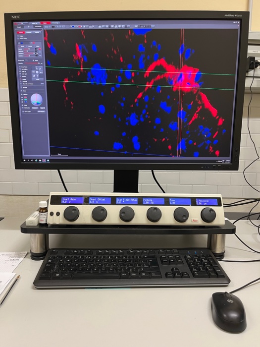
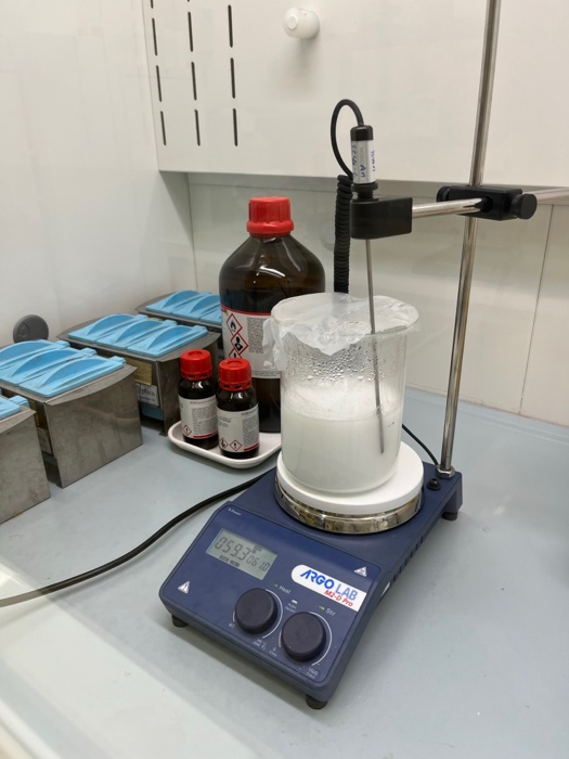
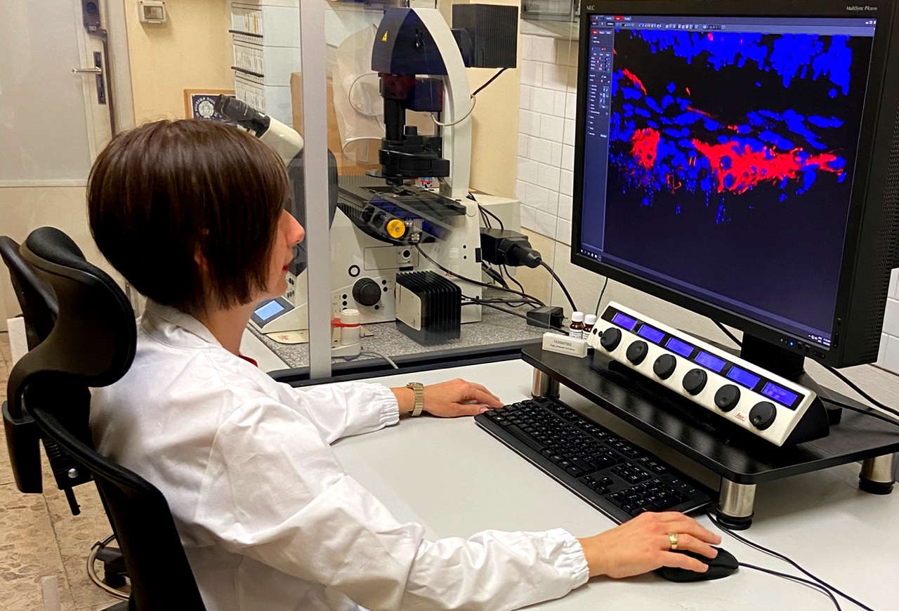
The Histology Unit of the University of Pisa has got Leica optical microscopes for the observation in brightfield and acquisition of representative images of histological, histochemical and immunohistochemical stainings performed mainly on fixed tissues, as well as a Leica TCS SP8 laser scanning confocal microscope, dedicated for observing and imaging of fixed and living samples.
Leica confocal platform is provided of systems for brightfield, polarized light, differential interference contrast (DIC) and fluorescence. In particular, the Leica TCS SP8 microscope is inverted and motorized in all components, has got dry and oil planapochromatic objectives. The scanning head supports 3 spectral photomultipliers of which one is Hybrid, with quantum efficiency greater than 40% at 500 nm and the ability to operate in single photon counting mode, and 1 photomultiplier for transmitted light, all independently adjustable. Up to 8 laser lines can be used simultaneously with separation of the signals in excitation and emission by means of an acousto-optical filter integrated in the scanning head and continuously adjustable, with selection windows of amplitude less than or equal to 2 nm and capable of switching times not exceeding 10 microseconds: 405 nm, Argon multiline (458, 476, 488, 496, 514 nm), 561 nm and 633nm lasers.
The Leica Application Suite (LAS) X software is set up for 3D imaging, spectral separation, colocation, and has been implemented with the lightning, to observe rapid biological processes in a precise and detailed manner with the benefits of ultra-fast imaging and super resolution up to 120 nm, navigator, to scanning large samples, and image analysis.
We have had the pleasure of weaving collaborations with many research groups of the University of Pisa for basic, translational and clinical studies, and we have given our support to local and international companies interested in deepening the effectiveness of phytotherapeutics on animal models.
Our experience, gained over the years, has allowed us to improve the techniques and implement the instruments, so as to be able to publish histochemical method works, as well as distribution and colocalization studies of cellular markers involved in important inflammation signaling.
PLoS One. 2015; 10(12): e0144630, 2015 Dec 16. doi: 10.1371/journal.pone.0144630 PMCID: PMC4682672
J Crohns Colitis. 2016 Oct;10(10):1194-204. doi: 10.1093/ecco-jcc/jjw076.
Br J Pharmacol. 2021 Oct;178(19):3924-3942. doi: 10.1111/bph.15532. Epub 2021 Jun 24.
Eur J Neurol. 2022 Oct 20. doi: 10.1111/ene.15607. Online ahead of print.
Cell cultues
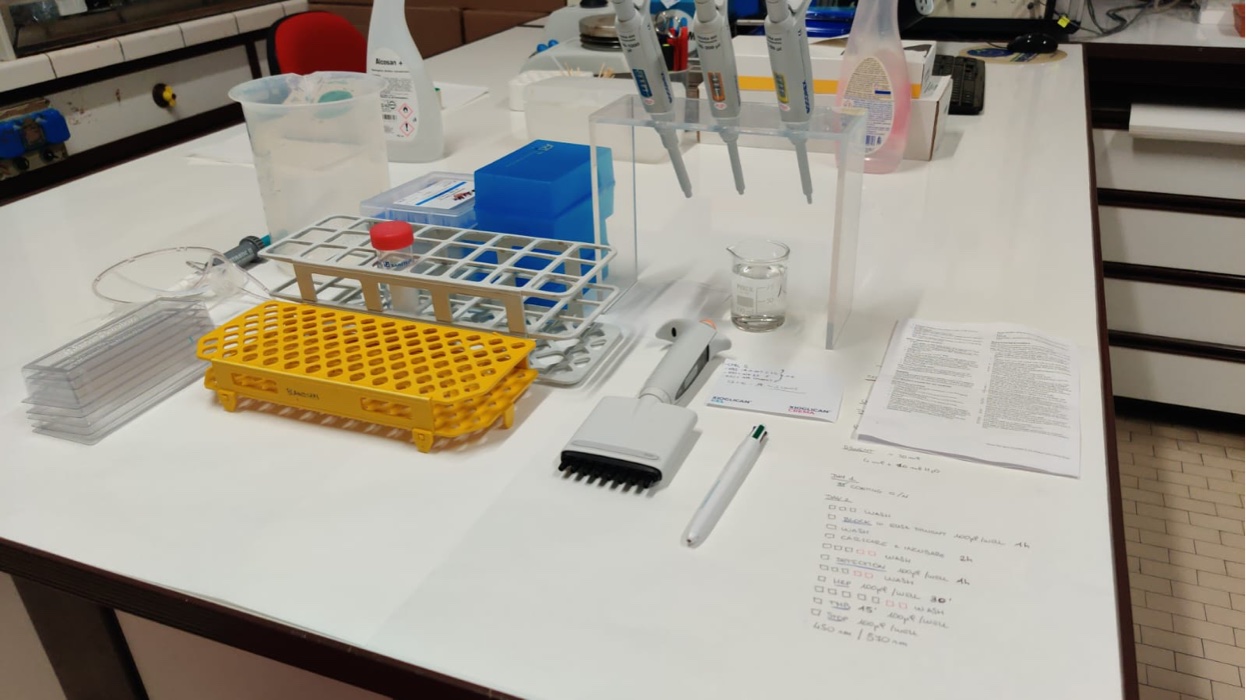
In our lab, we have facilities for cell cultures including a microbiological hood provided with a laminar flow workbench. We have several cell lines, including enteric glial cells (EGCs), intestinal epithelial cells (IEC-6, CaCo2) and immune cells (THP-1). Of note, we set up single cultures, co-cultures or 3D organoids to investigate the interactions between among cells resembling in vivo conditions
In addition, we are able to isolate a chosen cell type from fresh tissues using the technique of immunomagnetic cell sorting (“MACS”) and so consequently create a primary culture.
Functional studies
We have equipment for ex-vivo functional studies on isolated intestinal tissues. In particular, we study the
intestinal contractility on longitudinal and circular smooth muscles from ileal and colonic samples. Up to
four different specimens can be assessed in the organ baths. The preparations are connected to isometric
force transducers and challenged with electric stimuli in order to evaluate the contractility of the tissues. By
using specific antagonists or agonists, we can evaluate, in addition to the total electrical stimulation, also its
components, specifically isolated cholinergic and tachykininergic contractions.
Behavioural tests
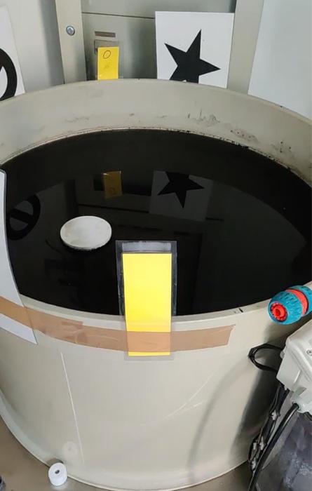
We have facilities in which we perform different behavioural tests on animal models. In the specific, our
most performed test is the Morris Water Maze, used to study spatial learning and memory. This test is used
in models of Alzheimer’s disease and Parkinsono’s disease to measure the effect of neurocognitive disorders on spatial learning and the
possible efficacy of treatments, to test the effect of brain damage in areas related to memory, and to study
how age influences cognitive function.

